Chapter 5 - Cartilage and Bone
Cartilage and Bone are specialized connective tissues that serve structural and supportive functions. Each is uniquely adapted to distinct mechanical demands. Cartilage is found in locations requiring flexibility, resilience, and shock absorption, such as joints, the ear, and the respiratory tract. In contrast, bone provides rigid support and resists deformation, forming the structural framework of the skeleton that protects organs, anchors muscles, and bears the body's weight.
CARTILAGE
Cartilage consists of three primary components: chondrocytes , extracellular fibers (collagen and/or elastic fibers), and a hydrated ground substance rich in proteoglycans and glycosaminoglycans. Its high-water content—up to 80% by weight—gives exceptional resistance to compressive forces, acting as a cushion and shock absorber. Meanwhile, the fibrous component provides tensile strength and elasticity, enabling cartilage to withstand stretching and return to its original shape after deformation.
Three Types of Cartilage:
- Hyaline Cartilage : Primarily type II collagen fibers embedded in abundant ground substance
- Elastic cartilage : Contains elastic fibers in addition to type II collagen, providing greater flexibility
- Fibrocartilage : Contains dense bundles of type I collagen along with type II collagen, providing exceptional tensile strength
Cartilage is avascular and its cells rely on diffusion for nutrients. Because of this, damaged cartilage heals poorly after injury.
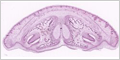
Hyaline Cartilage
Hyaline cartilage contains type-II collagen fibers and a highly hydrated ground substance. It is the most common cartilage and is found on articular surfaces, walls of the respiratory system (trachea and bronchi), and portions of the rib cage.
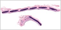
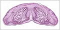
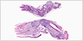
Elastic Cartilage
Elastic cartilage is similar to hyaline cartilage but also contains elastic fibers. It occurs where flexibility is required, such as the epiglottis, external ear, and auditory tubes.
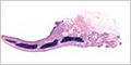
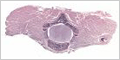
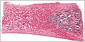
Fibrocartilage
Fibrocartilage contains a mixture of hyaline cartilage and dense regular connective tissue. It combines the tensile strength of collagen fibers with the resistance to compression of cartilage. It is found where tendons attach to bones, menisci and intervertebral discs.

CARTILAGE DEVELOPMENT
Chondrogenesis is the process by which cartilage is formed from condensed mesenchyme, differentiation into chondroblasts, and deposition of the extracellular matrix.
BONE
Bone serves three critical functions in the body: providing structural support, protecting vital organs, and acting as a calcium reservoir. The exceptional hardness and rigidity of bone result from mineralization of its extracellular matrix, where calcium phosphate crystals are deposited onto a collagen framework.
Morphologically, bone tissue exists in two distinct forms:
- Spongy bone (also called cancellous or trabecular bone): Porous, lattice-like structure found primarily at the ends of long bones and within vertebrae
- Compact bone (also called as cortical bone): dense outer shell of all bones and the shafts of long bones
Most bones in the human skeleton contain both compact and spongy bone tissue, strategically distributed to optimize strength while minimizing weight.
Unlike the avascular cartilage, bone maintains a rich vascular supply that supports its metabolic activity and remodeling capacity.

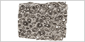
Spongy Bone
Spongy bone consists of a three-dimensional network of thin, interconnecting plates and rods called trabeculae. This porous structure is predominantly found in the interior of bones.
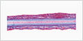
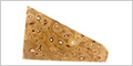
Compact Bone
Compact bone forms the dense, solid outer layer of all bones and comprises the entire shaft (diaphysis) of long bones. Its structure is highly organized into cylindrical units called osteons or Haversian systems , which align parallel to the long axis of the bone to resist bending and torsional forces. Each osteon consists of concentric rings of mineralized bone matrix called lamellae, arranged around a central Haversian canal that contains blood vessels and nerves.


BONE DEVELOPMENT
Osteogenesis is the process of bone formation and development. It involves cell migration, differentiation, extracellular matrix deposition, and mineralization.
Main Types:
- Intramembranous ossification - bone forms directly from mesenchymal tissue without a cartilage intermediate
- Endochondral ossification - bone develops by replacing a cartilage model
Key Cells Involved:
- Osteoblasts : Bone-forming cells that secrete bone matrix (osteoid)
- Osteocytes : mature bone cells embedded in the bone matrix
- Osteoclasts : Cells that break down and resorb bone tissue
- Chondrocytes : Cartilage cells (in endochondral ossification)
These cells also work together in a coordinated process called bone remodeling, which continues throughout life to maintain bone strength and repair damage.
Intramembranous Ossification
Intramembranous ossification forms bone directly from mesenchymal tissue without a cartilage intermediate. Mesenchymal cells differentiate directly into osteoblasts that produce the bone matrix ( osteoid ). During fetal development, this process forms the flat bones of the skull, the clavicles, and parts of the mandible.
Endochondral Ossification
Endochondral ossification forms bone by replacing a cartilage model through a complex sequence involving chondrocytes, osteoblasts, and calcification. This process forms most of the bones in the body, including long bones such as the femur and humerus.

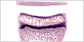
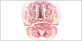
TOOTH DEVELOPMENT
Tooth development (odontogenesis) is the complex process by which teeth form from embryonic cells, grow, and erupt into the mouth.